5 MIN READ
Bacterial Leaf Streak in Corn
October 17, 2021
Bacterial leaf streak (BLS) was first documented in the United States in 2016, but symptoms were observed earlier in Nebraska in 2014. To date the disease has been found in Colorado, Illinois, Iowa, Kansas, Minnesota, Nebraska, Oklahoma, South Dakota and Texas. Bacterial leaf streak is caused by the bacterium Xanthomonas vasicola and has been found on field corn, popcorn, and sweet corn. The pathogen is a bacterium and once the plant is infected in the field, there is not currently a treatment available to protect other plants in the field. However, yield or quality losses have not yet been documented as a result of infection by BLS.
Epidemiology1,4
Currently it is believed that the BLS pathogen survives in previously infected plant residue. Secondary infections occur when bacterial exudates from infected plants are transported to healthy plants by wind, rain splash, or overhead irrigation water. Overhead irrigation when temperatures are above 90°F is associated with an increase in disease incidence. The pathogen penetrates corn leaves through the stomata, resulting in a typical banding appearance or pattern as lesions develop on the leaves. The pathogen appears to be restricted to areas between the main veins and does not invade vascular tissue. Because it is not systemic, it does not result in wilting like Goss’s wilt or Stewart’s wilt. While foliar lesions covering almost 40% of the leaf surface have been observed, no subsequent yield reduction was documented. Several weeds and other grass crops have been identified as host plants, including rice, oats, timothy, shattercane, Johnsongrass, green foxtail, yellow nudge sedge and downy brome.
Symptoms
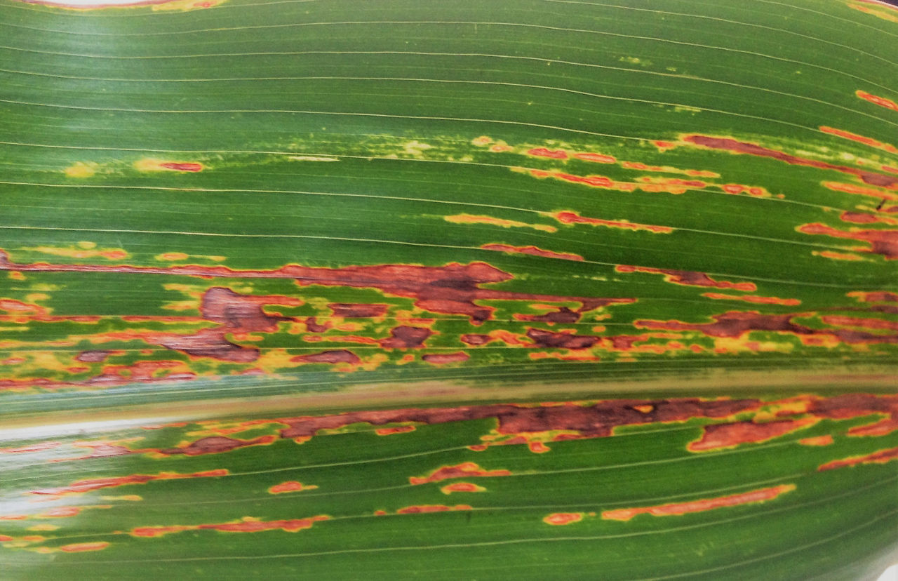
Symptoms can occur at any stage of corn growth but are typically observed first on lower leaves with spread to the middle and upper portion of the crop canopy after flowering. Initial symptoms appear as translucent, water-soaked streaks between veins (Figure 1) and progress to longer yellowish to necrotic streaks that can coalesce to form larger areas of symptomatic tissue (Figure 2). Bacterial exudates on the leaf surface may appear as small, dried, yellow-colored droplets. Lesions of BLS coalesce as leaf tissue dies creating large areas of necrosis.


Diseases with Similar Symptoms
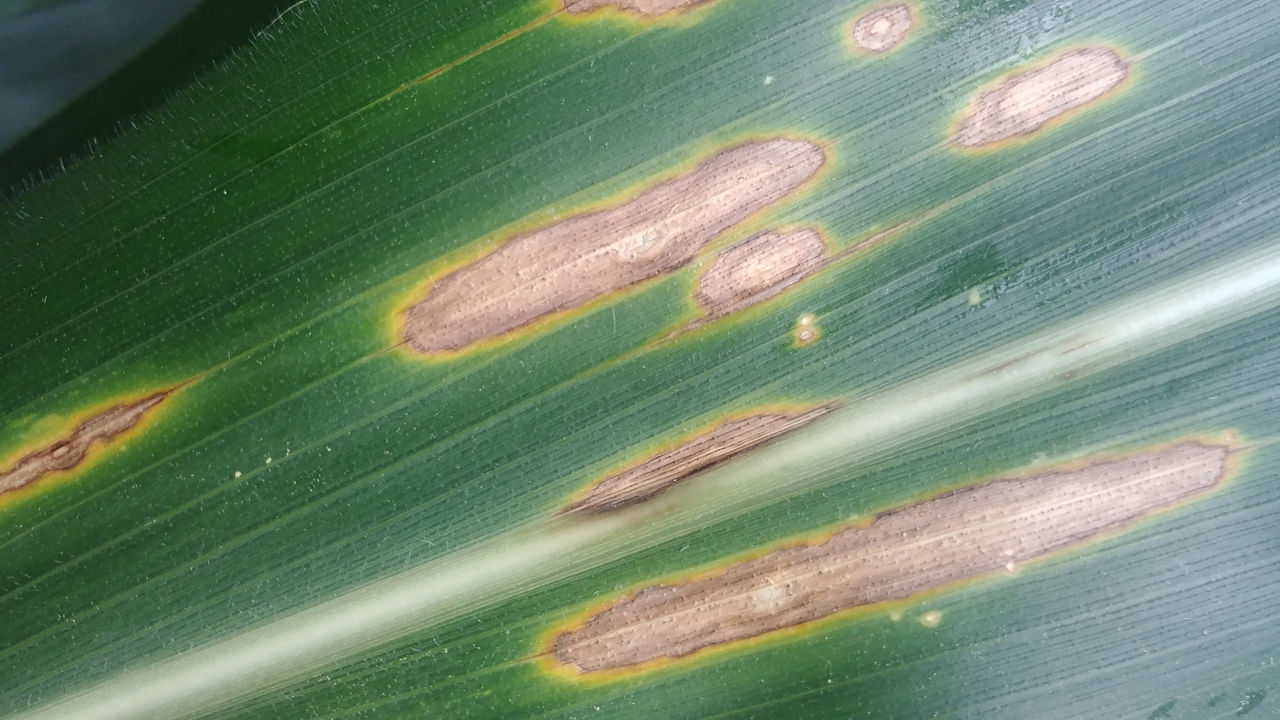
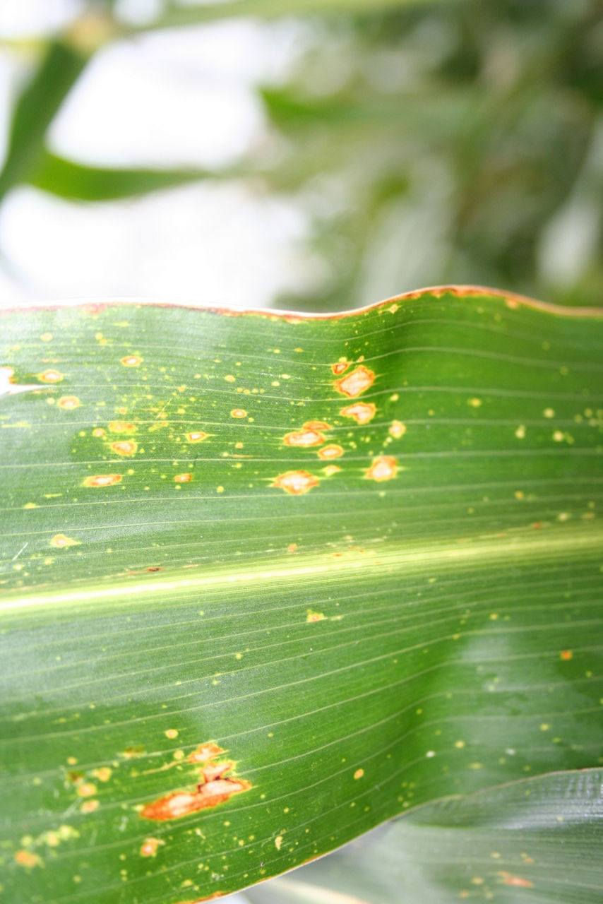
Gray Leaf Spot
Gray leaf spot (GLS) lesions look similar to BLS lesions; however, GLS lesions tend to be more rectangular with sharply defined edges when than BLS lesions while BLS lesion margins are wavy (Figure 3).

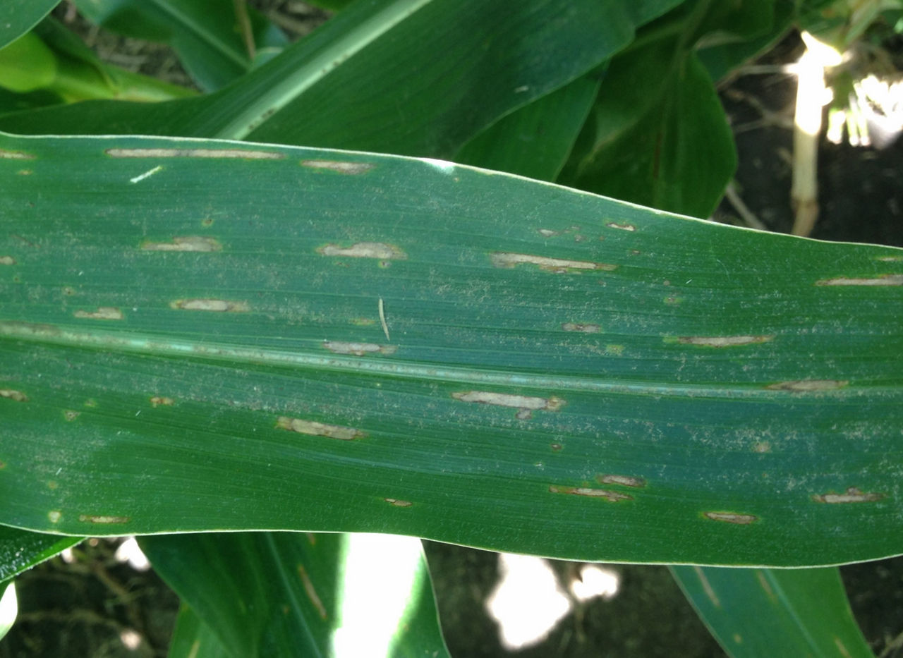
Figure 3. Bacterial leaf streak on top with wavy lined edged lesions, compared to gray leaf spot on bottom with straight edged lesions. Image courtesy of Emmanuel Byamukama, South Dakota State University.
When backlit, GLS lesions are more opaque with yellow margins, but BLS lesions are translucent (Figure 4).
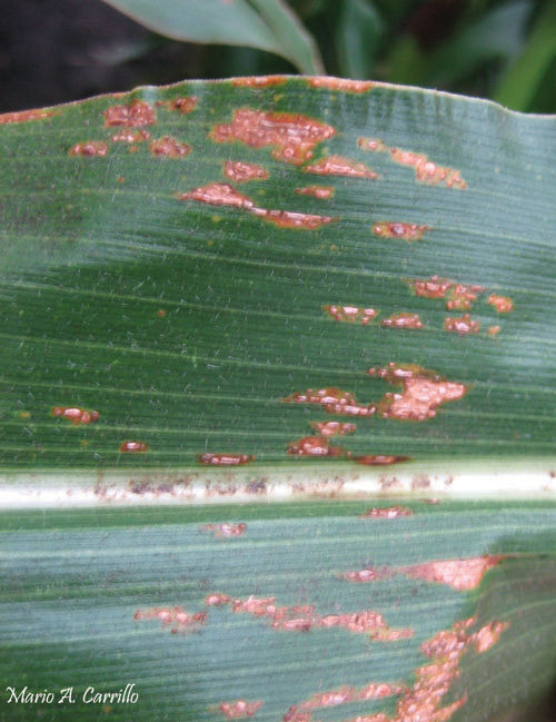
Common Rust
Common rust may be mistaken for BLS; however, common rust lesions are raised and blister-like (pustules) (Figure 5). When backlit, the pustules appear as dark circles.
Diplodia Leaf Streak
Early diplodia leaf streak (DLS) symptoms include dark, water-soaked, linear lesions that become tan with long, parallel sides and a bright yellow halo (Figure 6). As DLS lesions mature, they become more elliptical and look less like BLS. Additionally, small black spots (pycnidia) may appear in the center of mature lesions of DLS.
Southern Corn Leaf Blight
Mature southern corn leaf blight lesions can have brown to tan lesions with wavy sides and yellow halos in the lower canopy that resemble bacterial leaf streak (Figure 7). Southern corn leaf blight lesions are generally 1/8 to 1 inch long, but lesion length depends on corn product genetics.
Management1
Crop rotation to a non-host crop, such as soybean, wheat, or alfalfa.
Control weeds and volunteer corn as it can harbor the bacteria from year-to-year. Additionally, there are several grass weed hosts that may be alternative hosts.
Fall tillage can help speed the breakdown of crop residue resulting in lower inoculum levels; however, this control strategy must be balanced with risk of soil erosion.
Because BLS is caused by a bacteria, fungicides are not effective in managing this disease.
Sources
1 Jackson-Ziems, T. Bacterial leaf streak. University of Nebraska Extension. https://cropwatch.unl.edu/bacterial-leaf-streak.
2 Robertson, A. et al., 2016. Bacterial leaf streak of corn. Crop Protection Network. https://cropprotectionnetwork.org/resources/publications/bacterial-leaf-streak.
3 Sivits, S. and Jackson-Ziems, T. 2019. Bacterial leaf streak of corn in Nebraska. University of Nebraska Extension. https://cropwatch.unl.edu/2019/bacterial-leaf-streak-corn-nebraska.
5006_S5