5 MIN READ
Corn Diseases
July 29, 2020
What are Ear Molds and how do They Impact Yield and Grain Quality?
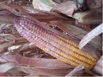
Gibberella Ear Rot (Figure 1) - This disease is a pink to reddish mold that starts at the ear tip and moves down the ear. Cool temperatures, below 72° F, with frequent rains at silking and during the two weeks after silking provide the best environmental conditions for this disease. This mold produces mycotoxins that can be toxic to livestock, particularly swine. This disease overwinters on corn debris and is more common in continuous corn fields compared to those in rotation.
Diplodia Ear Rot (Figure 2) - Diplodia is a gray mold which typically develops at the base of the ear. When scouting for this disease, the entire husk needs to be removed to determine the severity, which could be difficult if the husk has become stuck to the ear. This mold is usually found in continuous corn and reduced tillage scenarios. Warm, dry weather prior to silking followed by wet conditions after silking favors this mold. Diplodia can cause a reduction in yield and grain quality but does not produce mycotoxins; however, most livestock will not eat corn infected with this mold.
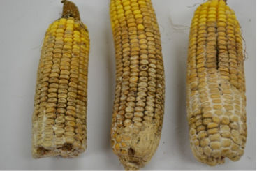
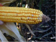
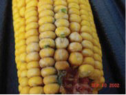
Fusarium Ear and Kernel Rot (Figures 3 and 4) - Fusarium can easily be distinguished by its brown coloration on the kernel caps or kernels with a white starburst pattern, individually or in groups across the ear. If the fungus is visible on the ear, the kernels appear white to salmon in color. This disease is more likely to become prominent with hot, dry weather during and after silking. It usually infects through damage from insects, hail, or mechanical means. Fumonisin, a mycotoxin associated with this ear rot, is toxic to livestock, particularly swine and equines.
How Do You Determine The Severity of an Ear Mold Problem?
To determine ear mold severity, the threshold can be determined by walking and counting the number of infected ears within a defined distance at several locations within the field. Proper harvesting, drying, and storage can help reduce the impact of these diseases. If ear molds are found, grain samples can be submitted to laboratories to determine if mycotoxins are present.
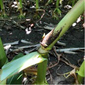
Anthracnose Stalk Rot vs. Purple Leaf Sheath vs. Physoderma Brown Spot and Node Rot
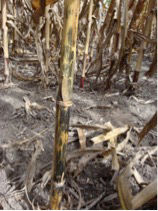
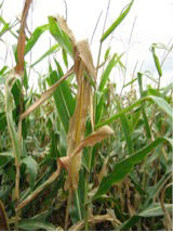
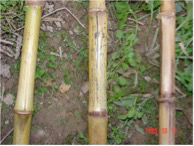
Anthracnose stalk rot is characterized by shiny black discoloration on the lower stalk (Figures 5 and 7). This disease can cause top dieback, with the elongated black lesions on the upper stalk under the upper leaf sheaths (Figure 6). The disease is most prevalent in continuous, reduced tillage corn scenarios. Early season rain and mid-season stress favor disease development and can be best detected about two weeks prior to physiological maturity. The presence of anthracnose leaf blight does not always result in stalk rot or top dieback later in the season and conversely, the lack of anthracnose leaf blight does not mean stalk rot or top dieback will not occur. Management options include the use of resistant products, tillage, and crop rotation.
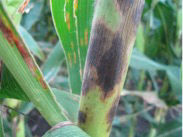
Purple leaf sheath, which usually appears after tasseling, should not be confused with the stalk discoloration of anthracnose stalk rot and Physoderma node rot. Purple leaf sheath is not a disease but a deterioration of leaf sheath tissue resulting from microorganisms feeding on pollen and dust particles that lodge between the sheath and stalk (Figure 8). The stalk remains healthy behind the discolored sheath.
Physoderma node rot can cause discoloration and breakage at the nodes (Figure 9). Brownish-orange sporangia are found at the rotted node and easily rubbed off. The node rot is more evident near maturity. The leaf lesions of Physoderma brown spot appear as very small round-to-oval shaped yellowish-brown lesions. Numerous lesions are evident and form bands across the leaf. Additionally, dark-purple to black spots develop on the leaf midrib. Physoderma is favored by early-season warm, wet, and humid conditions.
Jared Hauseman
Agronomist
Sources:
Diplodia ear rot. C.O.R.N. Newsletter. 2016-30. CFAES. Agronomic Crops Network. Ohio State University Extension. The Ohio State University. https://agcrops.osu.edu/.
Corn, soybean, wheat and alfalfa field guide, Bulletin 827. 2019. pg .44-47. Agronomic Crops Network. Ohio State University Extension. The Ohio State University.
Corn ear rots: Identification, quantification and testing for mycotoxins. 2016. Agronomic Crops Network. Ohio State University Extension. The Ohio State University. https://agcrops.osu.edu/.
Kleczewski, N. 2014. Anthracnose leaf blight and stalk rot of corn. Fact Sheets and Publications. University of Delaware. https://www.udel.edu/.
Robertson, A. 2016. Disease imposters. Integrated Crop Management. Iowa State University. https://crops.extension.iastate.edu/.
Websites verified 5/7/2020.
Performance may vary, from location to location and from year to year, as local growing, soil and weather conditions may vary. Growers should evaluate data from multiple locations and years whenever possible and should consider the impacts of these conditions on the grower’s fields. Channel® and the Arrow Design® and Seedsmanship At Work® are registered trademarks of Channel Bio, LLC. ©2020 Bayer Group. All rights reserved.